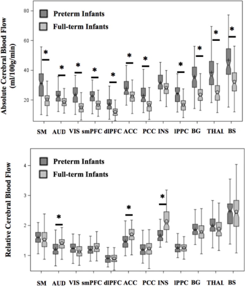
Figure 2. Unadjusted boxplots of regional cerebral blood flow assessed in the preterm (n=98) and full-term (n=104) population at term-equivalent age.
* Indicates the adjusted means between groups (preterm versus full-term infants) was significantly different (p<0.05) after adjustment for multiple comparisons using Tukey-Kramer, controlling for brain region, sex, gestational age at birth and day of life at MRI.
ACC = anterior cingulate cortex; AUD = auditory cortex; BG = basal ganglia; BS= brainstem; dlPFC = dorsolateral prefrontal cortex; INS = insula; lPPC = lateral posterior parietal cortex; PCC = posterior cingulate cortex; SM = sensorimotor cortex; smPFC = superior medial prefrontal cortex; THAL = thalamus; VIS = visual cortex.

