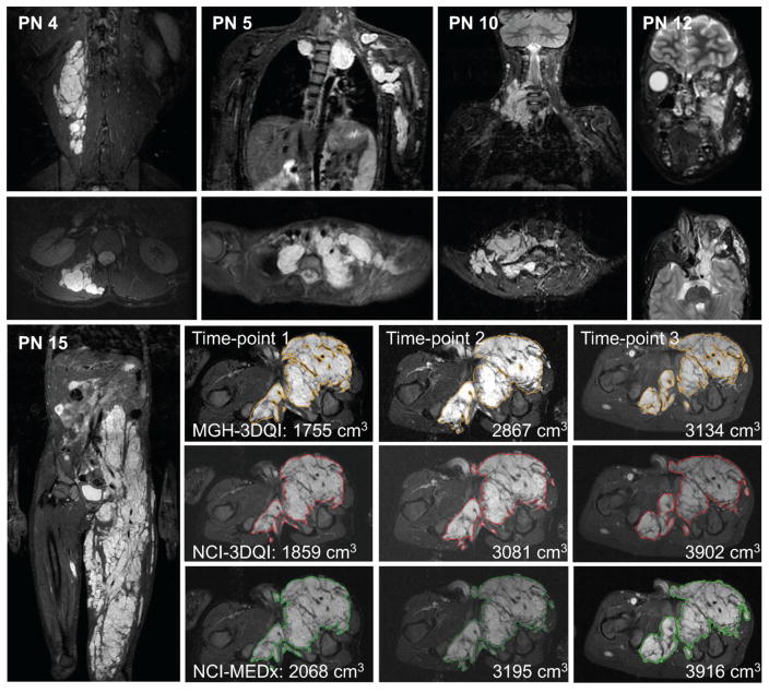
Figure 1. Examples of plexiform neurofibromas included in the study.
Coronal (top row), and axial (second row) STIR MR images of PN included in the study. PN 4: small, well defined PN in the right lumbar paraspinal muscle layer. PN 5: Large and highly complex PN in the upper chest, left shoulder, and arm. PN 10: medium sized right neck PN with moderately complex shape. PN 12: medium sized PN in left face, infiltrating the orbit, and facial muscles. PN 15: Coronal overview and segmentation contours at comparable levels in the axial plane from the three volumetric analyses, as labelled.

