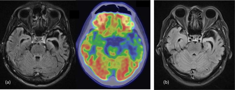
Figure 2.

Neuroimaging examples of case 1 and case 2. (a) case 1: MRI brain FLAIR images and FDG-PET brain images showed a predominant left anterior temporal lobe atrophy and hypometabolism. (b) case 2: MRI brain FLAIR images showed a predominant left anterior temporal lobe atrophy.
