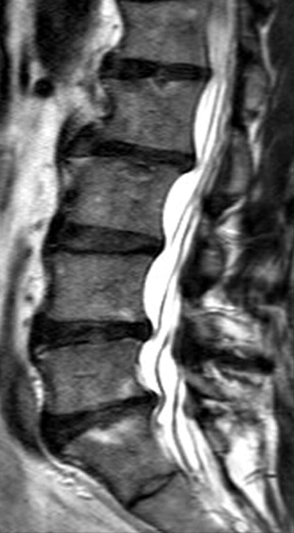
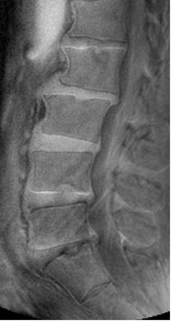
Figure 2.


Sagittal magnetic resonance imaging (MRI) of the lumbar spine of a subject with no chronic low back pain. (A) T2-weighted MRI noting multi-level disc degeneration and Modic changes. (B) Ultra-short time-to-echo (UTE) MRI noting no UTE Disc Sign (UDS).
