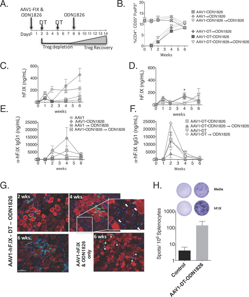
Figure 3. Combined effects of TLR9 stimulation and Treg depletion on systemic expression of and humoral immune responses after muscle-directed gene transfer.
(A) Timeline of experimental procedure for TLR9 stimulation and Treg depletion. The initial dose of the TLR9 agonist was combined with the vector and injected IM on day 0. Where indicated (⇒), the TLR9 agonist was injected in the same hind-leg on day 8. (B) Recovery of Tregs in peripheral blood as a function of time under various conditions. (C–F) Comparison of systemic hFIX antigen (C–D) and anti-hFIX IgG1 (E–F) levels in mice as a function of time after vector administration. (n=3–6 mice/group, *p=0.0178 AAV1-DT v. AAV1-DT-TLR9. (G) Immunofluorescent labeling of muscle tissue shows hFIX expression (red) and significant infiltration of CD8 cells (green) 2 weeks after AAV1-hFIX vector, TLR9 stimulation, and Treg depletion. (Arrows in insert point to centralized nuclei (blue) associated with new cell growth; ▲ indicate CD8+ (green) cells). (H) IFN-γ ELISpot showing a cellular immune response against hFIX in mice transduced with vector in the presence of ODN-1826 and Treg depletion, which was absent in mice treated with vector only (n=3–4/group; data are average ± SEM for epitope-stimulated minus mock-stimulated cultures).

