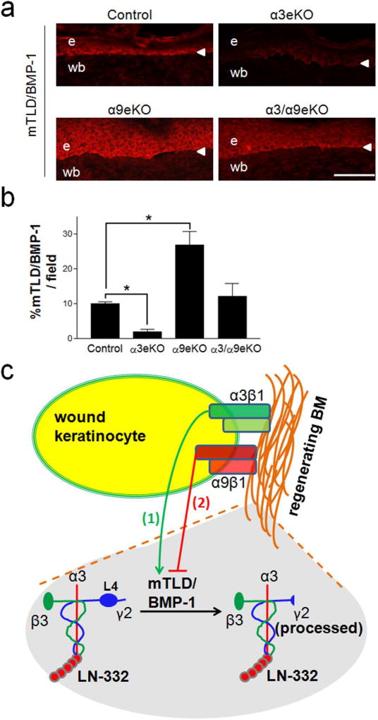
Figure 5.
α3eKO wound epidermis displays reduced mTLD/BMP-1 levels while α9eKO wound epidermis displays increased mTLD/BMP-1 levels. (a) Representative cryosections of 10-day reepithelialized excisional wounds were prepared from control, α3eKO, α9eKO, or α3/α9eKO mice and immunostained with anti-BMP-1. e, epidermis; wb, wound bed; arrowhead, BMZ. (b) mTLD/BMP-1 level was quantified for each genotype as percent positive staining per field of wounded skin; scale bar = 100 µm; mean ± SEM; n ≥ 5 mice per genotype; 1-way ANOVA followed by Newman-Keuls multiple comparison, *P<0.05. (c) Model illustrating our findings that (1) α3β1 promotes and (2) α9β1 inhibits expression of mTLD/BMP-1, thereby regulating LNγ2 processing in regenerating BM. Note that the L4 module (detected by anti-γ2L4m) is present only in unprocessed LNγ2 (left).

