
Figure 2.
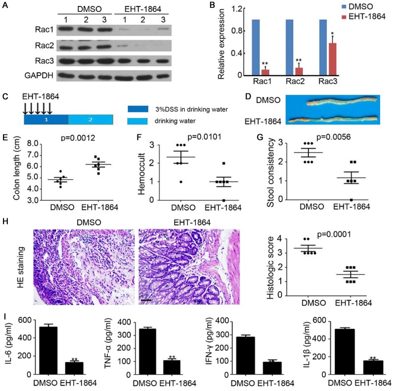
Inhibition of Rac impairs acute colitis in mice. A. Western blotting analysis of Rac1, Rac2 and Rac3 expression in colonic tissue of acute colitis mice. GAPDH was used as a loading control. B. The relative expression of Rac1, Rac2 and Rac3 was analyzed. n=3, **, P<0.01. C. Schema of acute colitis model and EHT-1864 treatment. D. The representative macroscopic photos of colon after DMSO or EHT-1864 injection in acute colitis mice. E. Colon length of acute colitis mice. n=6, **, P<0.01. F. Hemoccult of acute colitis mice. n=6, **, P<0.01. G. Stool consistency of acute colitis mice. n=6, **, P<0.01. H. HE staining of colonic tissue of acute colitis mice. The histologic score was calculated and analyzed. n=5, **, P<0.01. I. ELISA analysis of IL-6, TNF-α, INF-γ and IL-1β expression in colonic tissue of acute colitis mice. n=3, **, P<0.01.