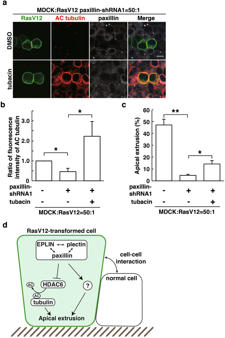
Figure 7.
Inhibition of HDAC6 partially rescues the paxillin-knockdown phenotype. (a) Effect of tubacin on accumulation of acetylated α-tubulin in paxillin-knockdown RasV12-transformed cells. MDCK-pTR GFP-RasV12 paxillin-shRNA1 cells were mixed with normal MDCK cells on collagen gels. Cells were fixed after 24 h incubation with tetracycline in the presence or absence of tubacin and stained with anti-acetylated α-tubulin (red) and anti-paxillin (grey) antibodies and Hoechst (blue). Scale bar, 10 μM. (b) Quantification of fluorescence intensity of acetylated α-tubulin. Fluorescence intensity of acetylated α-tubulin was analysed in each condition, and values are expressed as a ratio relative to paxillin-shRNA1 (-) tubacin (-). Data are mean ± SD from three independent experiments. *P < 0.05; n ≧ 30 cells for each experimental condition. (c) Quantification of the effect of tubacin on apical extrusion of RasV12 paxillin-shRNA1 cells. Apical extrusion was analysed after 24 h incubation with tetracycline. Data are mean ± SD from three independent experiments. *P < 0.05 and **P < 0.005; n ≧ 100 cells for each experimental condition. (d) The schematic of non-cell-autonomous changes in RasV12-transformed cells neighbouring normal epithelial cells.

