Figure 3.
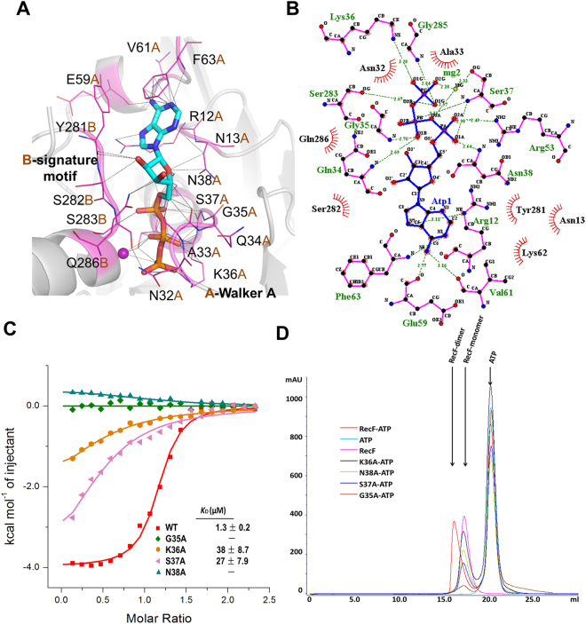
Walker A motif is essential to ATP binding and ATP-dependent dimerization. (A) ATP interacts with the residues in the Walker A motif and signature motif of RecF. All of the interacting residues are represented by sticks (magenta). (B) Extensive interactions between TTERecF and ATP. The plots were generated using LIGPLOT33. RecF residues and ATP are shown in pink and blue, respectively. H bonds are indicated by dashed lines (green). (C) ITC curves of TTERecF and the mutants of Walker A motif titrated into ATP. The ITC experiments involved 20 injections of 2 µL 1 mM ATP into 300 µL 70 µM RecF native or the mutants. (D) Size-exclusion chromatography analysis of TTERecF, TTERecF-ATP, ATP, G35A-ATP, K36A-ATP, S37A-ATP and N38A-ATP. Size exclusion chromatography was performed using a HiLord 16/60 Superdex 200 column (GE Health Life Sciences) at 0.5 ml/min in 50 mM Tris-HCl pH 7.0 and 300 mM NaCl. Protein elution was monitored by measuring the absorbance at 280 nm.

