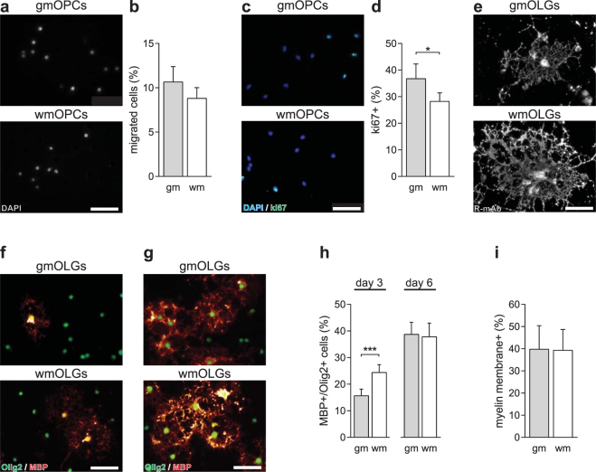
Figure 3.
WmOPCs proliferate less and differentiate faster than gmOPCs. Oligodendrocyte progenitor cells (OPCs) isolated from the cortex (gmOPCs) and non-cortex (wmOPCs) of neonatal rat forebrains were cultured for 4 hours (a,b) or 48 hours in the presence of PDGF-AA and FGF-2 (c–i), followed by differentiation for 3 (immature stage, e,f and h) or 6 days (mature stage, g–i). (a,b) OPC migration towards a 10 ng/ml PDGF-AA gradient (4 hours) was determined using a transwell assay. Representative images of migrated DAPI-stained OPCs are shown in a; quantitative analyses of the percentage of DAPI-stained migrated OPCs of the total number of plated cells in (b) (n = 10), (c,d) OPC proliferation was determined by immunocytochemistry for the proliferation marker ki67. Representative images are shown in (c), quantitative analyses of the number of ki67-positive of total DAPI-stained cells in (d) (n = 16, at least 150 cells analysed per independent experiment). Note the higher percentage of proliferating gmOPCs compared to wmOPCs. (e–i) OPC were differentiated for 3 (e,f, and h) and 6 days (g–i) and incubated with either (e) R-mAb, recognizing GalCer/sulfatide, or (f–i) double stained for MBP (red), a mature marker of oligodendrocytes (OLGs) and Olig2 (green), OLG lineage marker. Representative images are shown in (e,f) and (g); quantitative analyses of the number of MBP-positive OLGs of total Olig2-positive cells in (h) (n = 8 for 3 days, n = 10 for 6 days, at least 150 cells analysed per independent experiment) and the number of MBP-positive cells that elaborate myelin membranes in (I) (n = 10, 6 days). Note that after 3 days of differentiation wmOLGs are larger and morphologically more complex than gmOPCs. In addition, wmOPCs show an accelerated differentiation, while the number of MBP-positive cells bearing myelin membranes at day 6 is similar. Bars represent means. Error bars show the standard error of the mean. Statistical analyses were performed using a paired two-sided t-test (*p < 0.05, ***p < 0.001). Scale bar is 50 µm.

