Figure 8.
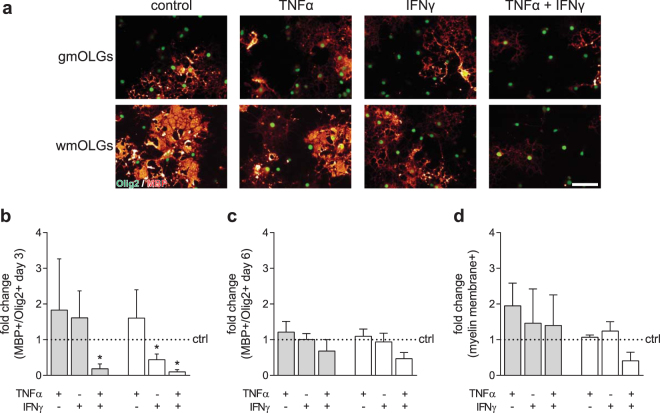
Exposure to IFNγ delays wmOPC, but not gmOPC differentiation. Oligodendrocyte progenitor cells (OPCs) isolated from the cortex (gmOPCs) and non-cortex (wmOPCs) of neonatal rat forebrains were left untreated or treated with 10 ng/ml TNFα, 500 U/ml IFNγ, or a combination of TNFα and IFNγ for 48 hours in the presence of PDGF-AA and FGF-2, followed by differentiation in the absence of cytokines. (a–d) OPC differentiation was determined at 3 (b) and 6 days (a,c,d) of differentiation using double staining for MBP (red), a mature marker of oligodendrocytes (OLGs) and Olig2 (green), an OLG lineage marker. Representative images at 6 days of differentiation are shown in (a); quantitative analyses of the number of MBP-positive cells of total Olig2-positive cells in (b) (3 days, n = 4) and (c) (6 days, n = 4) and the number of MBP-positive cells that elaborate myelin membranes in (d) (6 days, n = 4). Note that brief exposure to IFNγ at the OPC stage delays the differentiation of wmOPCs, but not of gmOPCs, while combined treatment with TNFα and IFNγ inhibited differentiation of either OPC. Grey bars represent gmOPCs, white bars represent wmOPCs (b,c,d). Error bars show the standard error of the mean. Bars represent mean relative to their respective untreated control, which was set at 1 for each independent experiment (horizontal line). Statistical analyses were performed using a one-sample t-test (*p < 0.05) to test for differences between treatments and their respective control and a one-way ANOVA with a Šidák post-test was used to test whether the response to TNFα, IFNγ and TNFα and IFNγ combined differed between gmOPCs and wmOPCs (not significant). Scale bar is 50 µm.

