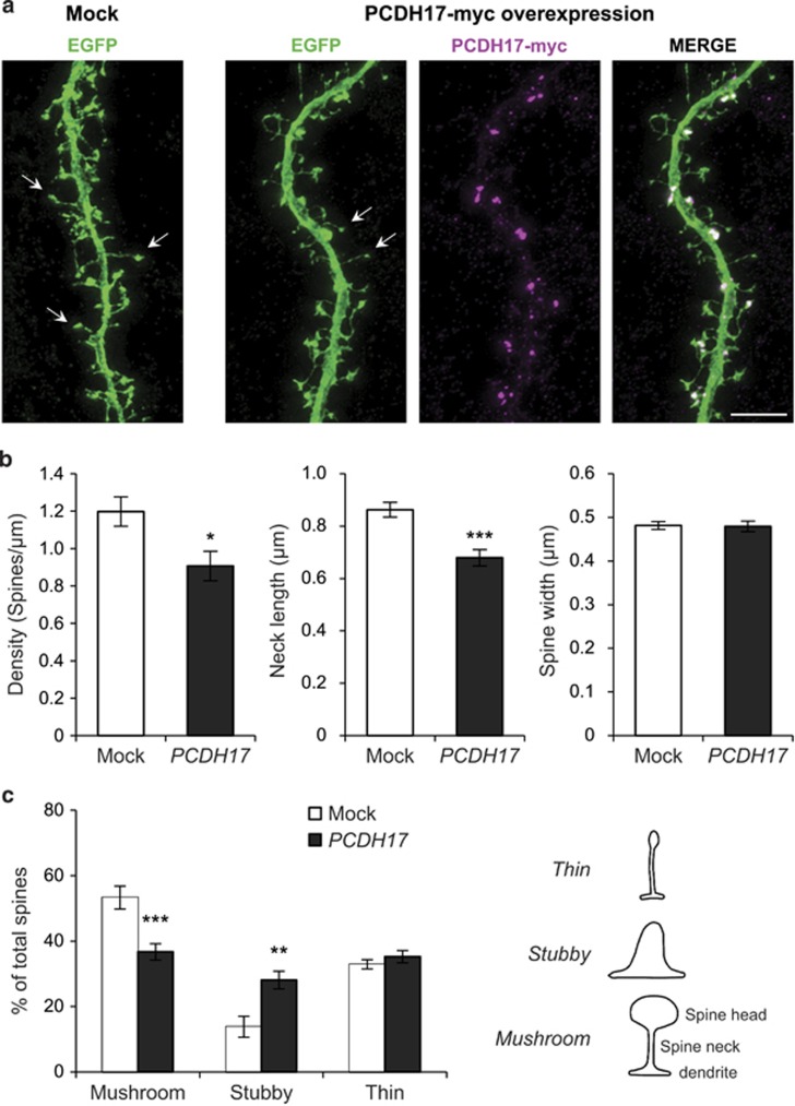
Figure 5.
Overexpression of PCDH17 decreases spine density and results in abnormal spine morphology in cultured cortical neurons. Scale bars represent 5 μm. (a) Cultured cortical neurons were transfected at DIV17-18 with EGFP plus mock plasmid or PCDH17-myc and maintained for additional one day. Neuronal morphologies were visualized by EGFP. Representative spines were arrowed in both groups. (b) Quantification of dendritic spine parameters (density, neck length and spine width). (c) Dendritic spines were divided in three different categories depending on their morphology: stubby, thin and mushroom, as indicated in the line drawing on the right. The diagram showed the percentage of total spines belonging to each category in Mock or PCDH17-Myc transfected cortical neurons. Dendritic spines were counted for each condition from four separate cultures. Error bars indicated s.e.m. *P<0.05, **P<0.005, ***P<0.001; Student's two-sided t-test (b) and two-way analysis of variance post hoc Bonferroni test (c).

