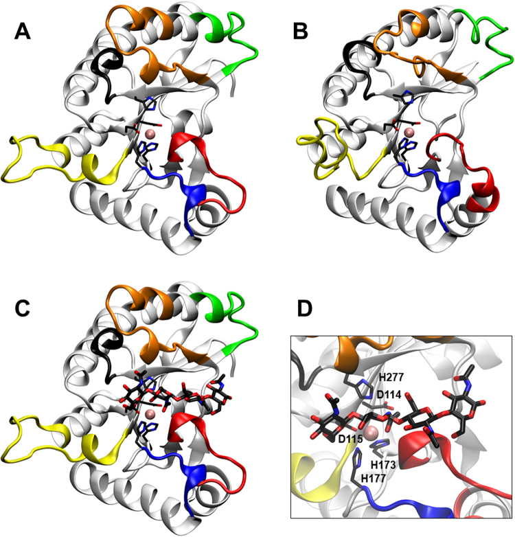
Figure 6.
Structural models of the PcCDA catalytic domain. (A) Model 1, using ClCDA (PDB 2IW0) and AnCDA (PDB 2Y8U) as templates. (B) Model 2, using ClCDA (PDB 2IW0) and VcCDA (PDB 4OUI) as templates. The loops are coloured as in Fig. 4 according to28. (C) Simulated docking of A4 ligand to Model 1, lowest energy binding mode which places the penultimate GlcNAc residue properly oriented for catalysis in subsite 0. (D) Magnification of the active site in Model 1, showing residues Asp115-His173-His177 (metal binding triad), Asp114 (general base), His277 (general acid), and the Zn2+ cation.

