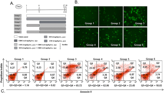
Figure 1.
(A) Graphical presentation of different groups of mice for in vivo chemoprotection study. (B) Effect of MUS on CBDCA-induced cytotoxicity and clastogenic effect in murine bone marrow cells. Photomicrographs after TUNEL assay were taken at 200× magnification. Brightly-stained nucleus represents an apoptotic cell, whereas, unstained cells represent non-apoptotic cells. (C) Inhibition of CBDCA-induced cell death of murine bone marrow cells by MUS. After treatment completion, murine bone marrow cells were isolated and stained with annexin-V and PI as described in Methods and analyzed by flow cytometry. The figure is a representative profile of at least three experiments in duplicate.

