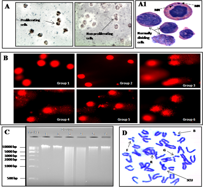
Figure 2.
Effect of MUS on CBDCA-induced cytotoxicity and clastogenic effect in murine bone marrow cells. (A) Representative photomicrograph (taken at 400 × magnification) of cell proliferation, where proliferating cells are BCIP/NBT stained and non-proliferating cells are unstained. (A1) Representative photomicrograph for the formation of micronuclei (indicated by broken arrow) after CBDCA treatment (1000X). (B) Effect of MUS on CBDCA-induced DNA damage in murine bone marrow cells. Photomicrographs after comet assay were taken at 400 × magnification. (C) Photomicrograph of agarose gel electrophoresis of genomic DNA. (D) Representative photomicrograph of chromosomal aberrations in a single metaphase plate taken at 1000 × magnification. Different anomalies like break [B], sister chromatid union [SCU], fragmentation [F], gap [G] and ring [R] are indicated.

