Figure 1.
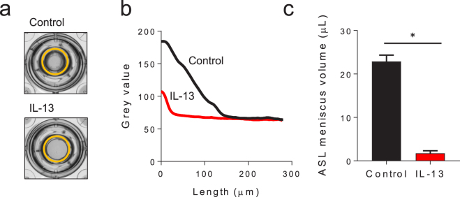
ASL meniscus volume is decreased in primary HBE cell culture following IL-13 exposure. Differentiated HBE cells were cultured ± IL-13 (10 ng/mL) for 3–5 days prior to measuring ASL meniscus volume. (a) Representative images of the apical surface of HBE cell cultures with an orange circle outlining the ASL meniscus. (b) Representative tracings of the change in light intensity through the apical meniscus used to measure the ASL meniscus volume in HBE cell cultures ± IL-13. (c) ASL meniscus volume in HBE cell cultures is decreased by IL-13. Data shown are mean ASL meniscus volume ± SEM of experimental replicates performed on cells from different 12 tissue donors, each replicate with 4–6 cultures, *p < 0.0001 by unpaired Student’s t-test. Control donor lines are represented in black, IL-13 treated donor lines are represented in red.

