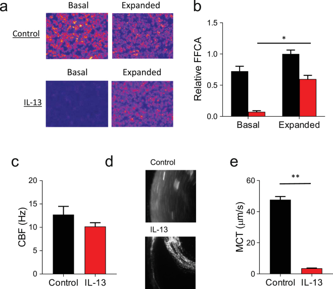
Figure 2.
IL-13 impairs mucociliary function in HBE. Control conditions are represented in black, IL-13 treated conditions are represented in red for all plots. (a) Combined serial representative images of the fraction of functional ciliary area (FFCA) of HBE cultures ± IL-13. Variations in light intensity representing ciliary motion are indicated in red and yellow. Images were obtained under basal conditions and following the expansion of the ASL with apical rinses of Ringer’s solution. (b) Relative FFCA is decreased by IL-13 under basal conditions and improved when the ASL is expanded. Data is expressed as mean FFCA ± SEM relative to control, n = 18 HBE cultures from 3 different tissue donors, *p < 0.0001 via paired Student’s t-test. (b) Ciliary beat frequency (CBF) in IL-13 treated HBE cell cultures approaches that of untreated controls after expansion with 10 µL Ringer’s solution. Data shown are mean CBF ± SEM (Hz), n = 3 experimental replicates from distinct tissue donors, each replicate is the mean CBF taken from >15 fields. (c) Representative images of mucociliary transit in HBE cultures with and without IL-13. (d) Mean mucociliary transit (MCT) is decreased by IL-13. Data shown are mean MCT ± SEM (µm/sec), n = 6 cultures each with >5 measured microspheres, **p < 0.0001 via unpaired Student’s t-test.

