Figure 10.
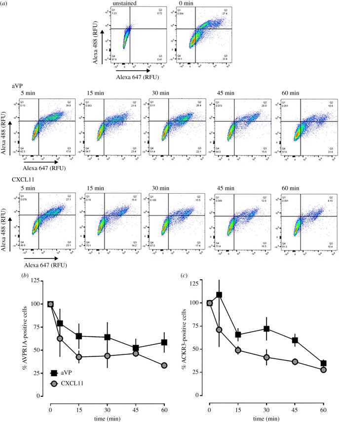
AVPR1A and ACKR3 co-internalize upon agonist stimulation in hVSMC. (a) hVSMCs were treated with 1 µM aVP or CXCL11 for up to 60 min, stained at 4°C with rabbit anti-AVPR1A/donkey anti-rabbit Alexa Fluor 647 and mouse anti-ACKR3/goat anti-mouse Alexa Fluor 488 and analysed for receptor expression via flow cytometry. RFU: relative fluorescence units. The horizontal and vertical lines show the gating thresholds for ACKR3 (Alexa 488) and AVPR1A (Alexa 647). (b) Quantification of AVPR1A-positive cells after incubation with aVP and CXCL11, as in (a). n = 3 independent experiments. (c) Quantification of ACKR3-positive cells after incubation with aVP and CXCL11, as in (a). n = 3 independent experiments.

