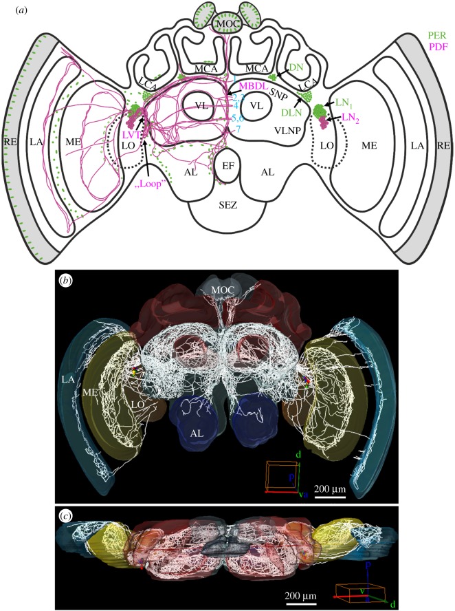
Figure 1.
PER- and PDF-positive cells in the brain of the honey bee A. mellifera. (a) Schematic presentation of the PER (green) and PER/PDF-positive cells (magenta) in the brain. The main fibre tracts of the PER/PDF-positive neurons (magenta) are additionally depicted in the left brain hemisphere together with the nuclei of PER-positive glia cells and photoreceptor cells (green). Abbreviations: 1–7, commissures 1–7; AL, antennal lobe; DLN, dorsolateral neurons; DN, dorsal neurons; EF, oesophageal foramen; LA, lamina; LCA, lateral calyx; LN1, lateral neurons 1; LN2, lateral neurons 2; LO, lobula; LVT, lobula valley tract; MBDL, median bundle; MCA, medial calyx; ME, medulla; MOC, medial ocellus; RE, retina; SEZ, suboesophageal zone; SNP, superior neuropils; VL, vertical lobe; VLNP, ventrolateral neuropils. (b,c) Reconstruction of all PDF fibres which emanate from the PER-/PDF-positive LN2 in a frontal (anterior) view (b) and from a dorsal view (c). Cell bodies are coloured, fibres are depicted in white. The PDF fibres invade all optic ganglia (LA, lamina; ME, medulla; LO, lobula), the ocelli (MOC, medial ocellus) and most parts of the bee's protocerebrum, as well as (sparsely) the ALs of the deutocerebrum. In addition, the tritocerebrum shows PDF-positive fibres around the oesophageal foramen that are hard to see in these reconstructions. Mushroom bodies and the central complex are free of PDF fibres.

