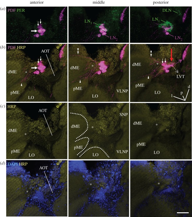
Figure 2.
Fibres arising from the PDF-positive somata towards the medulla and the protocerebrum. All pictures are from the same 60 µm-thick semi-frontal vibratome section that is tilted in the anterior–posterior as well as in the left–right plane to visualize the serpentine layer of the medulla (the orientation is indicated in the posterior plane of (b); orientation arrows: l, lateral; p, posterior; d, dorsal). Each picture shows an overlay of six confocal stacks in the anterior, middle and posterior area around PDF-positive somata, respectively. The vibratome section is labelled with anti-PER, anti-PDF, anti-HRP and DAPI. (a) PER and PDF labelling to demonstrate the vicinity of the LN1 and LN2 (compare figures 1a and 3). All LN2 cells contain PDF and PER. DLN dorsolateral clock neurons. PDF-positive neurites arising from the LN2 are hard to see, because we reduced PDF labelling intensity. The large arrow points to the largest PDF neuron, the small arrows to the smallest PDF neurons. (b) PDF labelling at higher intensity in order to reveal the arborizations of the LN2 on top of HRP labelling. HRP visualizes the major neuropil regions (plus the somata of neurons): dME, distal medulla; pME, proximal medulla; LO, lobula; SNP, superior neuropils; VLNP, ventrolateral neuropils. The PDF neurons send sparse neurites into a small neuropil reminding the accessory medulla of other insects (asterisks) and from there into the serpentine layer of the medulla (arrowhead) and to a minor degree into the most distal layer of the medulla (double arrowhead). In the direction towards the protocerebrum the PDF neurons send many fibres with dense varicosities onto the surface of the lobula (red arrow) and from there to the SNP (magenta broken arrow). From the densely labelled fibres anterior of the lobula, the lobula valley tract (LVT) originates and projects posteriorly and ventrally along the inner surface of the lobula (broken white arrow; for details on the LVT figure 4). Please note that the AOT is free of PDF fibres. (c) HRP labelling alone. The borders of the distal and proximal medulla (dME and pME) and the LO are indicated by broken white lines in the middle picture. The serpentine layer, in which the PDF neurons project (see (b)) is located between the dME and pME. (d) DAPI and HRP labelling. The neuropils marked by HRP are free of somata and nuclei. Large tracts such as the AOT usually contain the nuclei of glia cells, whereas the putative accessory medulla (asterisk) is basically free of nuclei. Scale bar, 50 µm.

