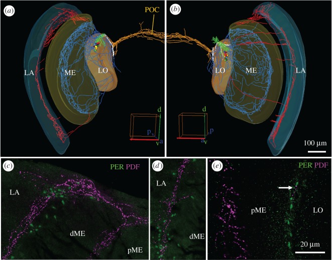
Figure 5.
PDF fibres in the medulla and lamina. The upper row shows reconstructions of both optic lobes. (a) PDF fibres running to the lamina (LA) are mainly present in the dorsal medulla (ME) and to a lesser extent in the ventral ME. (b) The fibres running to the LA are also most prominent in the dorsal optic lobe, but in addition, fibres projection in an anterior fan over the distal surface of the ME to the LA is more frequent. For better distinction, PDF fibres invading the LA are shown in red, those invading the ME in blue, those in the dense arborization area in front of the lobula (LO) in green, those in the lobula valley tract in white and those in the posterior optic commissure (POC) in orange. (c–e) Magnifications of PER/PDF double labelled vibratome sections in the optic lobes. (c) Dorsal part of the LA and ME (overlay of six confocal stacks). Note that the fibres entering the proximal LA are in close vicinity to PER-positive glia cells. The ME is divided into proximal (pME) and distal (dME) part, which are separated by PDF-positive fibres in the serpentine layer. In the serpentine layer only few PER-positive glia cells are located. (d) The PDF fibres in the more ventral part of the LA are also accompanied by PER-positive glia cells (overlay of six confocal stacks). (e) PER-positive putative glia cells between ME and LO (overlay of three confocal stacks). In this region only sparse PDF innervations are found (white arrow).

