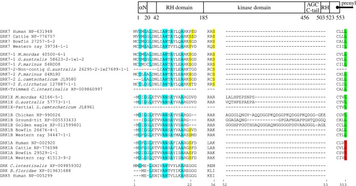
Figure 10.
Comparison of residues between visual GRK isoforms for a selection of taxa and positions. Schematic at top shows overall organization of the GRK1/GRK7 protein molecule. All numbering is for human GRK7. Cyan highlighting indicates the presence of the expected residue at the seven sites implicated in the binding of recoverin/S-modulin. Yellow highlighting indicates the Ser residues (at sites 22/23 and 36) that are subject to phosphorylation by PKA. The next segment shows the insertions (found immediately after the residue corresponding to 52 in human GRK7) in lamprey GRK1Xs and avian GRK1Bs. At the far right, green highlighting denotes that the terminal residue of the ‘CaaX’ prenylation motif is Leu (which provides the signal for geranylgeranylation), whereas red denotes a Ser or Ala (which provides the signal for farensylation). The entire alignment for all GRK sequences is shown in electronic supplementary material, table S2, and modelled molecular structures are presented in electronic supplementary material, figure S7.

