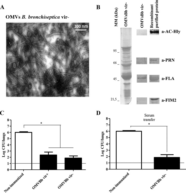
FIG 5.
(A) Transmission electron microscopic image of negatively stained OMVs obtained from B. bronchiseptica 9.73 in the avirulent phase (OMVBbvir−) (scale bar, 200 nm). (B) Analysis of OMVBbvir− by 12.5% (wt/vol) SDS-PAGE, with visualization with Coomassie brilliant blue. Molecular masses are indicated on the left. On the right, immunoblots of OMVBbvir− and the purified recombinant proteins AC-Hly, PRN, flagellin (FLA), and FIM2, as detected with the respective specific polyclonal mouse antibodies, are shown. (C) Effect of active i.p. immunization with OMVBbvir− in the mouse intranasal challenge model. (D) Effect of passive immunization with sera collected from OMVBbvir−-immunized mice. For panels C and D, B. bronchiseptica 9.73 was used as the challenge bacteria (1 × 10⁶ CFU, in 40 μl). Three biological replicates were performed, with the results from a representative one being presented. The data represent the means for 4 mice per group, at 7 days after challenge. The horizontal lines indicate the lower limit of detection. The numbers of bacteria recovered from the mouse lungs, expressed as the mean ± SEM (error bars) of log10 CFU in the lungs, are plotted on the ordinates for the lung samples from the mice receiving the immunogen, in the form of vesicles (C) or serum (D), indicated on the abscissas. *, significant differences (P < 0.001).

