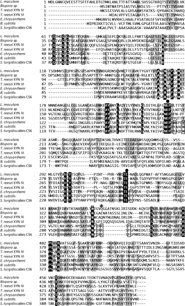
FIG 1.
Comparison of amino acid sequences of GH30 xylanases from S. turgidiscabies C56, E. chrysanthemi, B. subtilis, T. reesei XYN VI, T. reesei XYN IV, Bispora sp. strain MEY-1, and Leptosphaeria maculans. Amino acid sequences were aligned by using ClustalW software (37). Identical amino acids are shown in black and gray boxes. ●, catalytic residue; *, conserved arginine in bacterial GH30 enzymes. Amino acid residues located at cleft region conserved in bacterial GH30 enzymes are boxed.

