
Figure 1.
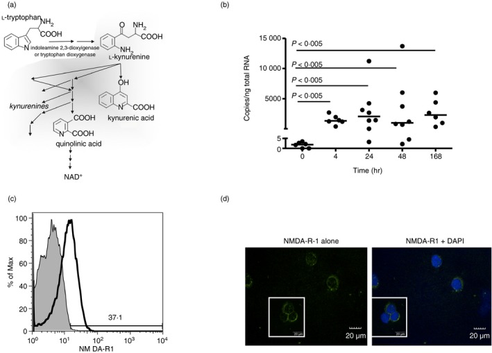
Anti‐CD3/CD28 activation induces elevated N‐methyl‐d‐aspartate receptor 1 (NMDA‐R1) expression by peripheral CD4+ T cells. (a) Scheme of kynurenine pathway in tryptophan catabolism. (b) Freshly isolated CD4+ T cells, activated with anti‐CD3/CD28 antibody for the indicated times, were examined for NMDA‐R1 expression by quantitative PCR (n = 9 for detailed kinetics) with medians (Mann–Whitney P‐values) shown. (c) Flow cytometric analysis of NMDA‐R1 protein expression on fresh CD3+/CD4+ primary T‐cell populations that were resting or stimulated with anti‐CD3/CD28 for 24 hr. Data are from one individual, representative of eight individuals examined. (d) Confocal laser‐scanning immunofluorescence microscopy of NMDA‐R1 expression at 24 hr on anti‐CD3/CD28 activated primary CD4+ T cells. NMDA‐R1 (green); DAPI (blue, right). Data are from one individual, representative of three examined.