
Figure 1.
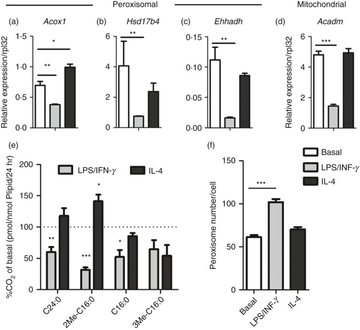
Both peroxisomal and mitochondrial β‐oxidation are strongly reduced in classically activated macrophages. (a–d) Gene expression of peroxisomal acyl‐CoA oxidase 1 (Acox1) (a), multifunctional protein‐2 (Hsd17b4) (b) and enoyl‐CoA, hydratase/3‐hydroxyacyl CoA dehydrogenase (Ehhadh) (c) and mitochondrial medium chain acyl‐CoA dehydrogenase (Acadm) (d) in classically (LPS/IFN‐γ) and alternatively (IL‐4) activated bone‐marrow‐derived macrophages (BMDM) compared with basal conditions (n = 4 versus 4 versus 4); (e) Degradation of substrates by peroxisomal β‐oxidation (2‐Me‐C16:0 and C24:0), mitochondrial β‐oxidation (C16:0) and peroxisomal α‐oxidation (3Me C16:0) in classically and alternatively activated BMDM (n = 4 versus 4 versus 4) expressed as percentage of CO 2 release in basal conditions; (f) Number of peroxisomes per cell in classically and alternatively activated BMDM compared with basal conditions (n > 15). Bars represent mean ± SEM. Statistical differences based on one‐way analysis of variance test: *P < 0·05, **P < 0·01, ***P < 0·001.