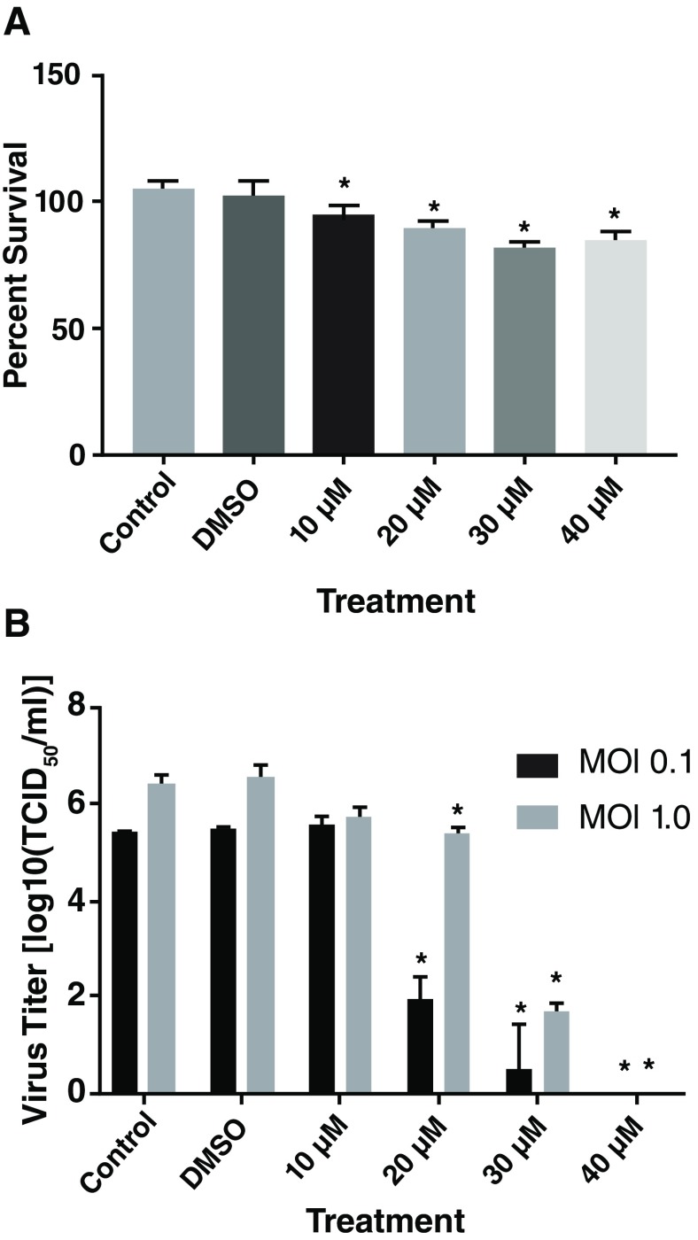
Figure 3. Effect of MF on hPDF cell proliferation and CHIKV181/25 replication.
( A) hPDF cells were treated with increasing doses of MF and MTT assay was done to determine the proliferation of cells. Since, MF was dissolved in dimethyl sulfoxide (DMSO), a DMSO concentration equivalent to concentration in 40µM dose was used in the assay. The control was saline treated cells. Significant effects on cell proliferation was observed in MF treated group compared to saline treated control groups. ( B) hPDF Cells were treated 4–6 h with MF and infected with CHIKV181/25 with either MOI 0.1 or 1. Virus titer in the cell supernatants was measured at 24 h post infection. Turkey’s multiple comparison test using 2 way ANOVA was performed to determine significance (*P < 0.05). Values are presented as ± SD. Values are presented as ± SD. * P ≤ 0.05 vs control group ( A & B), and ^ P <0.05 between two MOIs ( B). Data is representative of two repeat experiments.

