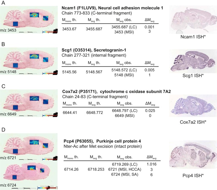
Fig. 5.
MALDI-MS images of top-down identified proteoforms of Ncam1 C-terminal fragment (A), Scg1 internal fragment (B), Cox7a2 C-terminal fragment (C) and intact Pcp4 N-terminally acetylated after initiator methionine excision (D) with their corresponding top-down (LC) and MSI m/z and their in situ hybridization images (ISH*). All values in tables are given in a.m.u. See also supplemental Data S7. *Image credit: Allen Institute (47).

