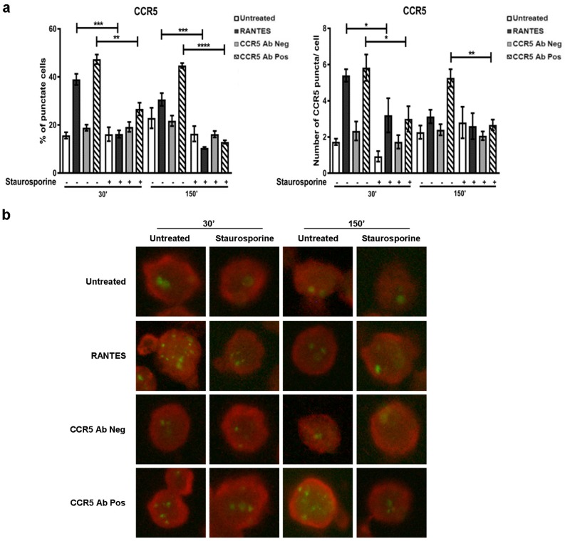
Figure 1.
CCR5 internalization in the presence of a pan-kinase inhibitor (staurosporine). After 1 h of pre-treated with staurosporine (50 nM), R5-SupT1-L23 cells were stimulated, or not, with CCR5 Ab Pos, RANTES (positive control) and CCR5 Ab Neg (negative control). The cells were harvested at 30 min and at 150 min (30 min incubation, wash, additional 120 min incubation in medium without stimuli). (a) The percentage of cells with the punctate form of CCR5 (left) and the number of CCR5 puncta per cell (right) in cells treated or not with the stimuli, with or without staurosporine treatment, stained with anti-CKR5(D6), are stated. Data are representative of three independent experiments. Bar graphs represented mean ± standard deviation (SD) of three independent experiments. Student’s t-test was performed and p-values are shown. * p ≤ 0.05, ** p ≤ 0.01, *** p ≤ 0.001, **** p ≤ 0.0001; (b) a representative immunofluorescence image of cells positive for CCR5 detection (lens magnification: 63×). Evans Blue dye was used as a counter stain.

