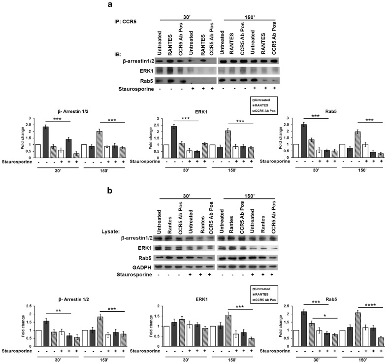
Figure 2.
Role of the phosphorylation status in the CCR5 signalosome formation. After 1 h of pre-treated with staurosporine (50 nM), R5-SupT1-M10 cells were stimulated, or not, with CCR5 Ab Pos, RANTES (positive control). The cells were harvested at 30 min and at 150 min (30 min incubation, wash, additional 120 min incubation in medium without stimuli). (a) Co-IP on cell lysates was performed for CCR5 followed by immunoblots for β-arrestin1/2, ERK1 and Rab5 expression; (b) western blot for β-arrestin1/2, ERK1 and Rab5 in total cell lysates was accomplished. Band density was determined with the TINA software (version 2.10, Raytest, Straubenhardt, Germany), and it is shown as fold change over a housekeeping gene. Bar graphs represented mean ± SD of three independent experiments. Student’s t-test was performed and p-values are shown. * p ≤ 0.05, ** p ≤ 0.01, *** p ≤ 0.001, **** p ≤ 0.0001. Data are representative of three independent experiments.

