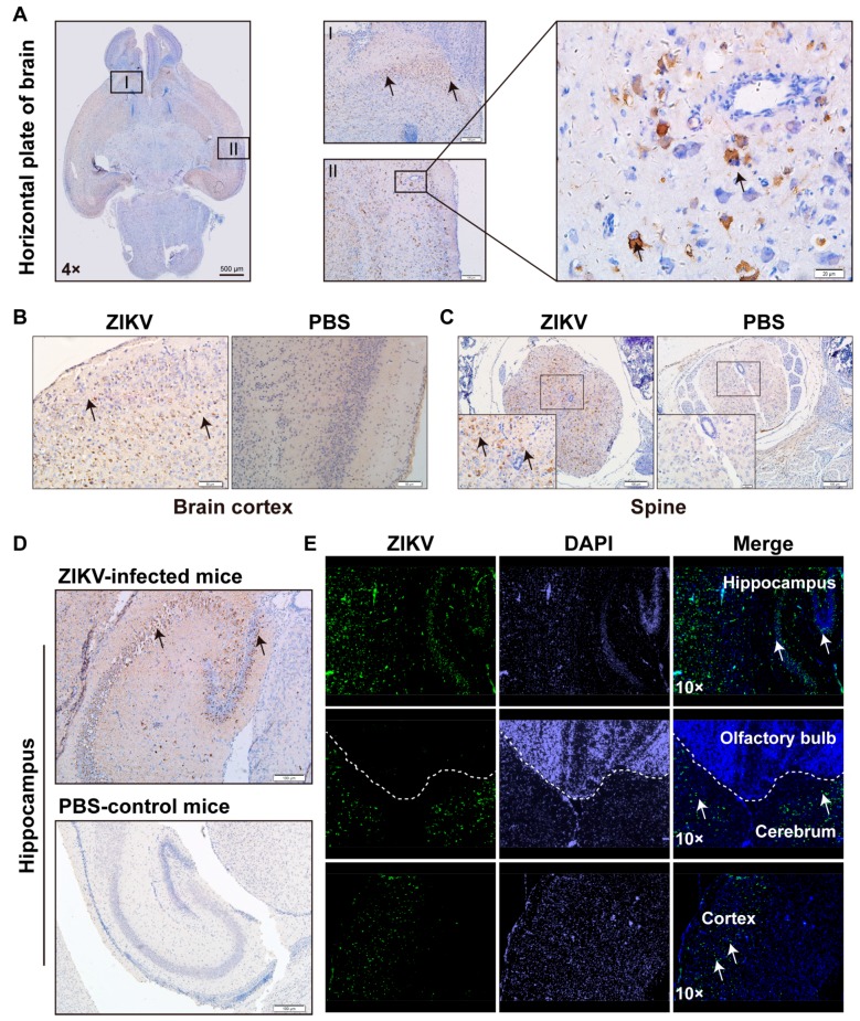
Figure 5.
ZIKV infected multiple areas of CNS. (A) Horizontal section of the brain of the ZIKV-infected mouse was stained with anti-E protein. The area I and II was amplified to show the infection of ZIKV (brown color stand for the positive infection; (B–D) Immunohistochemistry (IHC) analysis of CNS tissues after being infected with PRVABC59. The 1-day-old C57BL/6 mice were infected i.p. with 106 TCID50 PRVABC59 per mouse or PBS. Mice were sacrificed at 11 dpi to isolate brain tissue for sectioning and staining by the IHC method. Representative pictures from PRVABC59-infected brain tissue slides were selected to show (B) brain cortex; (C) spine; (D) hippocampus; (E) the same as in (B–D) but stained with IFA for hippocampus, olfactory bulb, cerebrum, and cortex. Scale bars: A left 500 μm, A middle, 100 μm, A right, 20 μm; B 50 μm; C 100 μm; D 100 μm.

