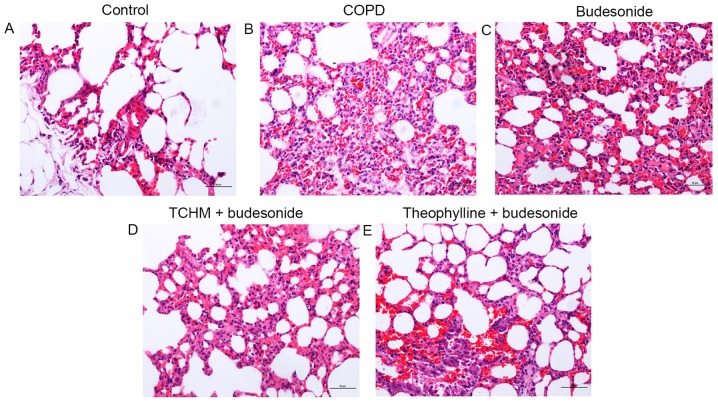
Figure 1.
Histopathological images. Lung tissue slices from each treatment group were stained with hematoxylin and eosin and photographic images were captured under a microscope. Magnification, ×200. (A) Control group, (B) COPD group, (C) budesonide group, (D) TCHM + budesonide group and (E) theophylline + budesonide group. One representative image from each group is shown (n=8). COPD, chronic obstructive pulmonary disease; TCHM, traditional Chinese herbal medicine.

