
Figure 5. Thymic ECs can be cultivated ex vivo and mediate enhanced thymic regeneration upon exogenous administration after damage.
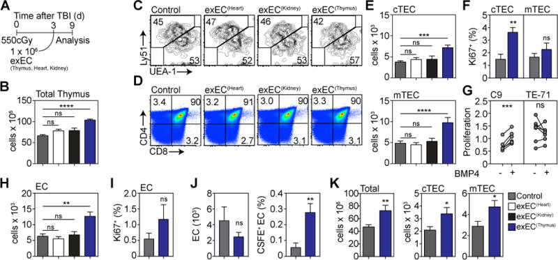
ECs were FACS sorted from the thymus (n=25), heart (n=10) or kidney (n=10) based on expression of VE-cadherin and transduced with the viral gene E4ORF1. These cells are referred to as exEC. In order to model immune injury we exposed 6–8 week-old C57BL/6 mice to TBI and 1 × 10⁶ exEC were administered iv at day 3 after TBI (n=25 in control group). (A) Experiment schematic. (B) Total thymic cellularity at day 9 after TBI. (C–D) Concatenated flow cytometric plots detailing the proportion of (C) TECs and (D) thymocyte subsets. (E) Absolute number of cortical and medullary thymic epithelial cells (cTEC and mTEC respectively). (F) TEC proliferation was measured on day 3 by Ki-67 staining. (G) C9 or TE-71 cells were stimulated with BMP4 (100ng/ml) for 24 hours after which proliferation was assessed (n=6–7 independent experiments). (H) Absolute number of ECs. (I) EC proliferation on day 3 after exEC administration. (J) exECs were generated and labeled with CFSE and 10 × 106 cells were transferred on day 3 after TBI. 4 hours after transfer CFSE expression was assessed by VE-Cad+ cells in the thymus. Displayed are total EC number and proportion of CFSE+ ECs (n=13–15 from three independent experiments). (K) Total thymic cellularity and absolute number of cTEC and mTEC 28 days after TBI and administration of 1 × 10⁶ thymus-derived exEC on day 3 (n=10/group). Graphs represent mean ± SEM of at least 2 independent experiments. *, p<0.05; **, p<0.01, ***, p<0.001.