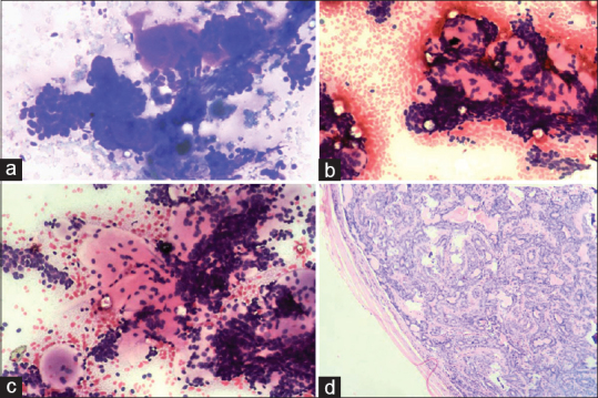
Figure 1.

(a-c) Photomicrograph of cytology of basal cell adenoma exhibiting clusters and cords of basaloid cells with variable amount basement membrane like stromal material and palisading of tumor cells at periphery and also in the basement membrane material (a: Leishman and Giemsa stain, ×40 b-c: H and E stain, ×40). (d) Photomicrograph showing histology of basal cell adenoma, the tumor was composed of bland basaloid cells separated by many abundant amorphous hyaline membranous stroma (H and E stain, ×100)
