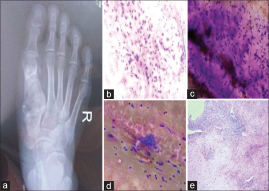
Figure 1.

(a) Radiograph of the right foot showed a lytic lesion in the head of the first metatarsal bone. (b) Smears were moderately cellular with round to ovoid cells embedded in a chondromyxoid matrix (H and E, ×100). (c) Stellate cells and spindle-shaped fibroblast-like cells were also seen (MGG stain, ×100). (d) Occasional scattered multinucleated giant cells were observed (MGG stain, 100×). (e) Biopsy showed pseudolobules of myxoid and chondroid tissue separated by zones of fibrous tissue (H and E, ×40)
