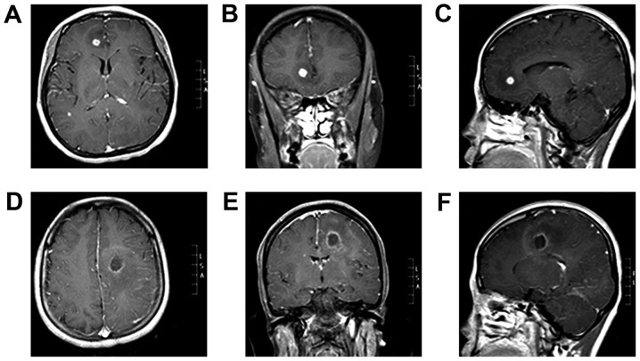
Figure 1.
Initial magnetic resonance imaging scan indicating two brain abscess lesions. (A-C) Right frontal lobe lesions (size: 8×8×8 mm, volume 0.2 cm3). (D-F) Left temporal lobe lesions (size: 19×19×20 mm, volume 2.9 cm3). (A and D) Axial contrast-enhanced T1WI. (B and E) Coronal contrast-enhanced T1WI. (C and F) Sagittal contrast-enhanced T1WI. T1W1, T1 weighted image.

