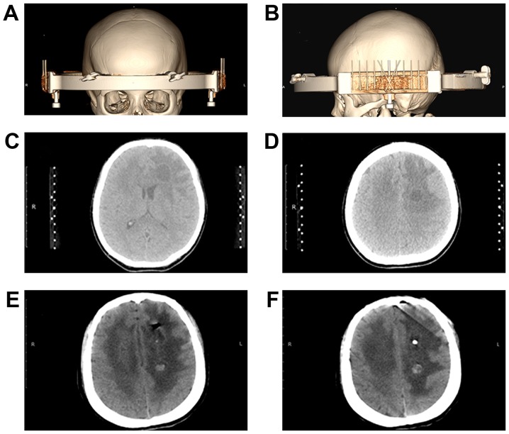
Figure 3.
3D CT images of stereotactic biopsy and cyst fluid aspiration surgery. (A) Stereotactic surgery localization and an axial image of 3D CT reconstruction. (B) Stereotactic surgery localization and sagittal image of 3D CT reconstruction. (C) Stereotactic surgery targeting the left frontal lesion selection. (D) Stereotactic surgery targeting the left semi-oval center lesion. (E) CT scan of the head conducted 1 day following stereotactic biopsy indicating a decrease in the size of the abscess in the left frontal cavity and a shadow of gas present in the brain parenchyma. (F) CT scan of the head conducted 1 day following stereotactic biopsy indicating the drainage tube positioned in the center of the abscess. The abscess in the left semi-oval center was notably smaller. A round and high-density shadow was present in the cavity. CT, computer topography; 3D, three-dimensional.

