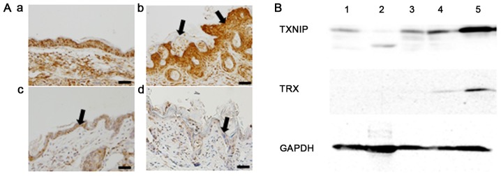
Figure 5.
TNF-α, TXNIP and TRX expression after the treatment. Histological features of the skin region. Three weeks after the initial treatment, skin specimens were obtained and fixed with 4% PFA. To observe inflammatory changes, TNF-α antibody was used. (Aa) Normal, saline-treated skin was included as a control (bar, 50 µm). (Ab) Hyperkeratosis and epidermal thickening (arrows) were observed after radiotherapy, with strong TNF-α staining. (Ac) Weak TNF-α staining was observed with D-allose treatment (arrow). (Ad) Radiation-induced epidermal thickening and TNF-α staining were reduced with additional D-allose treatment (arrow). (B) Western blot analysis of TXNIP and TRX expression. Proteins were obtained from: 1, normal skin with saline treatment; 2, normal skin with D-allose treatment for 2 weeks; 3, tumor tissue with saline treatment; 4, tumor tissue with D-allose treatment for 2 weeks; and 5, tumor tissue with D-allose treatment for 3 weeks. TXNIP, thioredoxin interacting protein; TRX, thioredoxin; TNF-α, tumor necrosis factor-α.

