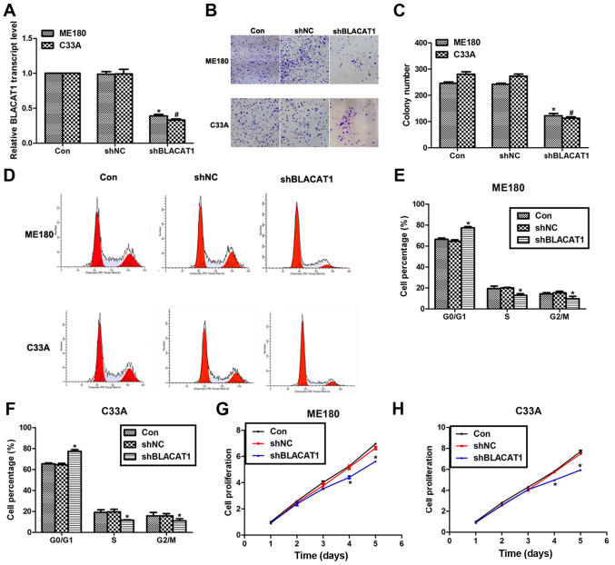
Figure 2.
Knockdown of BLACAT1 inhibited cell proliferation in human cervical cancer in vitro. (A) RT-PCR analysis was performed to assess the expression of BLACAT1 in ME180 and C33A cells upon transfection of shBLACAT1. (B) Representative images of colony formation assays for ME180 and C33A cells. (C) Colony formation assay was performed to assess the formed colonies of ME180 and C33A cells upon transfection of shBLACAT1. *P<0.05, vs. Con in ME180 cells. #P<0.05, vs. Con in C33A cells. (D) Representative images of cell cycle assays for ME180 and C33A cells. (E) Cell cycle analysis showed the cell percentage in each phase upon shBLACAT1 transfection in ME180 cells. (F) Cell cycle analysis showed the cell percentage in each phase upon shBLACAT1 transfection in C33A cells. (G) Cell proliferation assay was performed to reveal the cell proliferative rate of ME180 cells upon shBLACAT1 transfection in a continuous five days. (H) Cell proliferation assay was performed to reveal the cell proliferative rate of C33A cells upon shBLACAT1 transfection in a continuous five days. *P<0.05, vs. Con.

