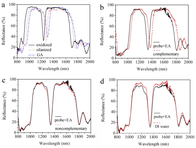
Figure 5.
(a) Reflectance spectra of double Bragg mirror structure following (nL1nH1)9(nL2nH2)9 sequence after oxidization, after silanization and after glutaraldehyde (GA). (b) Resonance shift of the double Bragg mirrors for 10 μM complementary deoxyribonucleic acid (DNA). Negligible resonance shift for (c) 10 μM non-complementary DNA and (d) Deionized water (DI).

