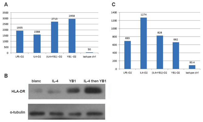
Figure 3.

M2 macrophages exhibited increased HLA-DR and decreased CD206 expression levels. (A) HLA-DR expression levels in M2 macrophages activated using LPS, IL-4, YB1, a combination of YB1 and IL-4 or an isotype. (B) HLA-DR expression confirmed using western blotting. (C) CD206 expression levels in M2 macrophages activated using LPS, IL-4, YB1, a combination of YB1 and IL-4 or an isotype. HLA-DR, human leukocyte antigen-antigen D related; CD206, mannose receptor; LPS, lipopolysaccharide; IL, interleukin; YB1, Salmonella typhimurium strain YB1.
