Figure 1.
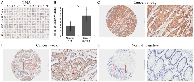
Immunohistochemical staining for FMO5 in colon cancer and normal colon tissues. (A) Staining pattern of the TMA section. (B) Immunoreactive score in cancerous tissues was higher than in normal colon tissues (IRS: Normal: 3.50±1.69 vs. cancer: 7.24±2.20, P=0.000). **P<0.01. (C) Images showing a strong FMO5 staining in the cytoplasm of tumor cells. The C is enlarged images of L04 spot. (D) FMO5 expression was weak in the cytoplasm of cancer cells. The D is enlarged images of K04 spot. (E) FMO5 expression was negative in the normal colon tissues. The E is enlarged images of L18 spot. The red arrows in C and D show positively stained cells. Original magnifications, ×100 and ×400. FMO5, flavin-containing monooxygenase 5.

