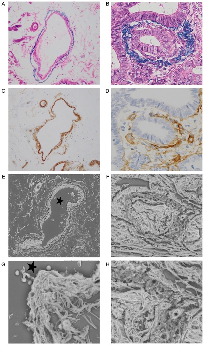
Figure 3.
Histological findings of (A and B) hematoxylin and eosin, Victoria Blue B and (C and D) collagen IV staining, and (E-H) three-dimensional ultrastructural findings in LV-SEM. Panels A, C, E and G show a normal vein in serial sections, while panels B, D, F and H show a vein invaded by tumor cells with positive matrix metalloproteinase-7 expression in serial sections. (A) Victoria Blue B staining was specifically visible in the elastic fibers of the adventitia (magnification, ×200). (B) Victoria Blue B staining of adventitia was thinner on the invasion side (magnification, ×400). (C) Collagen IV staining was visible in the intima. Collagen IV of the normal vein was stained as a strong and thick linear pattern (magnification, ×200). (D) Collagen IV staining of the intima was weak and interrupted by the invading tumor cells (magnification, ×200). (E) Serial section stained with phosphotungstic acid (Masson's trichrome staining) and assessed by LV-SEM (magnification, ×300). (F) Veins invaded by tumor cells were characterized by an irregular change in the two vascular layers in LV-SEM (magnification, ×800). (G) A normal vein was characterized by a two-layered structure with smooth elastic fibers of the adventitia and a mesh-like structure of collagen fibers in the intima by LV-SEM (magnification, ×1,500). (H) The thin portion of collagen IV staining was characterized by disappearance of the intimal mesh-like structure of collagen fibers in LV-SEM (magnification, ×1,500). The invading tumor cells in an isolated and clustered form were visible within the altered venous walls in LV-SEM (magnification, ×1,500). The stars in (E) and (G) indicate the same lumen. LV-SEM, low vacuum-scanning electron microscopy.

