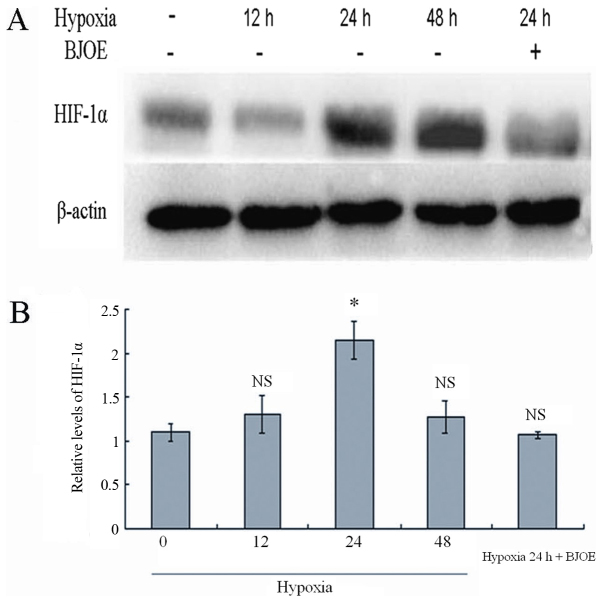
Figure 5.
(A) Western blot analysis for HIF-1α in ECA109. (B) Semi-quantitative levels of HIF-1α in ECA109. ECA109 cells were treated with BJOE under hypoxic conditions for 24 h. The control cells were cultured without BJOE under normoxic, and hypoxic conditions for 12, 24, and 48 h. Subsequently, proteins were extracted from cells. A total of 40 µg protein were loaded. The experiments were performed according to the procedures aforementioned in the materials and methods section. Comparable to normal protein levels were detected after 12 h under hypoxic conditions. However, HIF-1α levels increased to the maximum in ECA109 cells after 24 h under hypoxic conditions. In response to treatment with 5 mg/ml BJOE for 24 h under hypoxic conditions, the HIF-1α protein levels in ECA109 cells were notably inhibited by BJOE. Data are shown as mean ± standard deviation. *P<0.05, compared with the control; NS, no significance, compared with the control. HIF-1α, hypoxia-inducible factor 1α; BJOE, Brucea javanica oil emulsion.

