Figure 3.
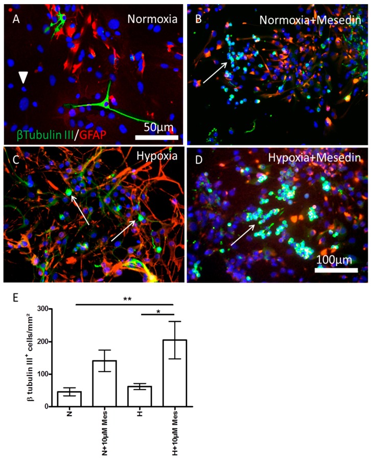
Neuroprotective effect of mesedin on DIV7 APC. The number of β-tubulin III-positive neurons (green) was assessed by double staining with glial fibrillary acidic protein (GFAP, red) in normoxic control (A), mesedin treated C57BL/6 APC upon normoxia (B), hypoxic control (C), and mesedin treated C57BL/6 APC upon hypoxia (D). Scale bar in (A): 50 µM, in (B–D): 100 µm. Arrowhead in A indicate the population of β-tubulin III and GFAP negative cells. Arrows in (B–D) indicate β-tubulin III-positive neurons/neuronal precursors. (E) Quantification of β-tubulin III-positive neurons/neuronal precursors upon normoxic (N) and hypoxic (H) culture conditions with 10 µM mesedin (N + Mes or H + Mes) in comparison to the respective controls (N vs. H). The cells were quantified from n = 5 coverslips, and normalized to mm2. The cell nuclei are counterstained with 4′,6-diamidine-2′-phenylindole dihydrochloride (DAPI, blue). Data are presented as mean ± SEM, and analyzed by two-way ANOVA with Bonferroni’s comparison test. * p < 0.05; ** p < 0.01.

