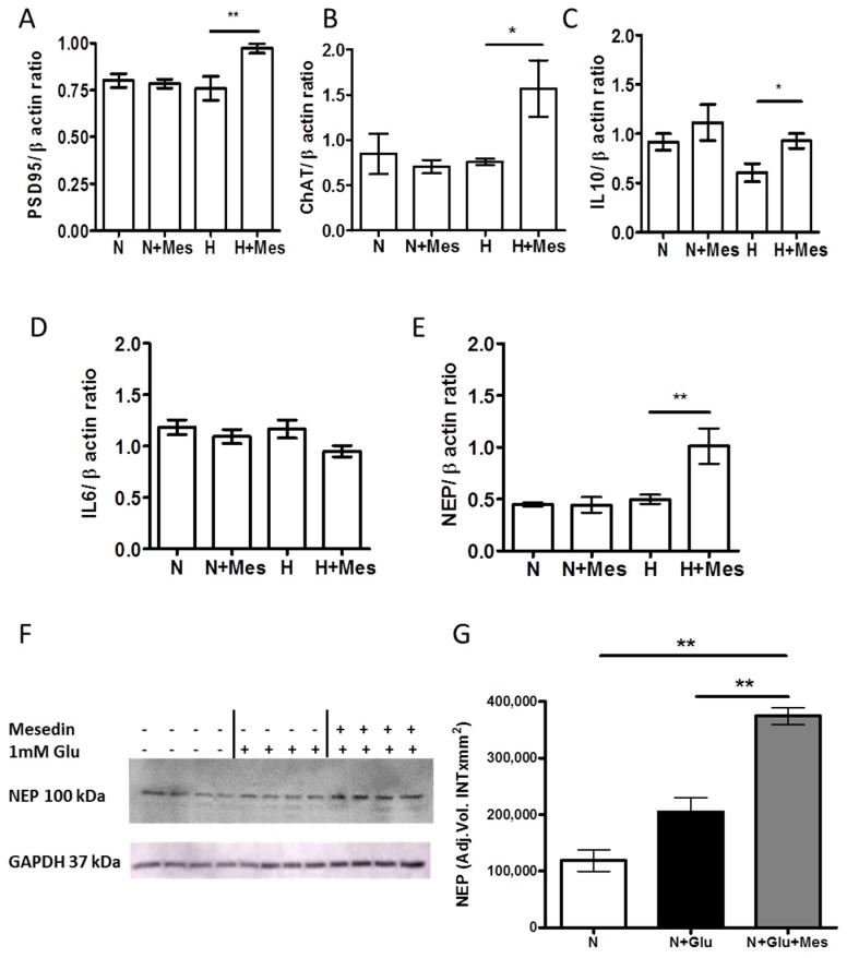
Figure 5.
The impact of mesedin on the cholinergic, synaptic, and inflammatory markers in wild type APC and Aβ degrading function of 3×Tg-AD APC. (A–E) qPCR analyses of postsynaptic density protein 95, PSD95(A), choline acetyltransferase, ChAT (B), Interleukine-10 (C), Interleukine-6, IL-6 (D), and neprilysin, NEP (E) upon normoxia (N), hypoxia (H) with 10 µM mesedin (N + Mes; H + Mes) vs. respective controls (N; H). (F) Representative Western blot and (G) densitometric analysis of NEP in APC from two-month-old 3×Tg-AD mice upon normoxia (N) with and without glutamate (Glu), and 10 µM mesedin (n = 4). Glycerinaldehyd-3-phosphat-Dehydrogenase (GAPDH) served as loading control. Data are presented as mean ± SEM and analyzed by one-way ANOVA with Bonferroni’s comparison test. * p < 0.05; ** p < 0.01.

