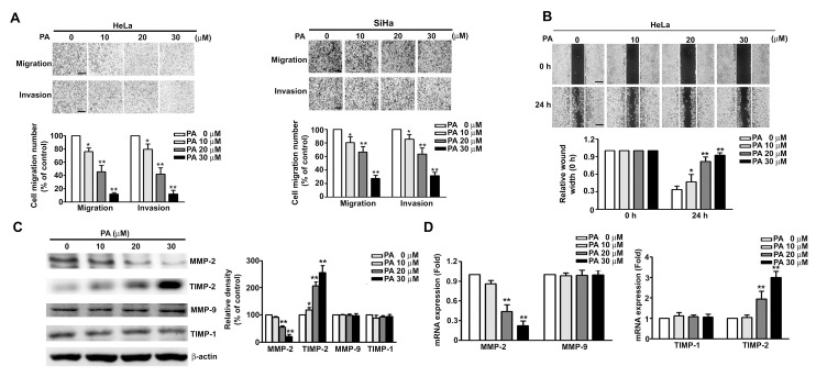
Figure 3.
Effect of PA on cell migration/invasion, wound closure, and protein expression of MMPs and TIMPs in SiHa and HeLa cells. (A,B) Cells were treated with various concentrations of PA (0 to 30 μM) for 24 h, followed by measurement of cell migration and invasion and relative wound width. (C,D) Cells were treated as above, then harvested for measurement of MMP-2, MMP-9, TIMP-1, TIMP-2 proteins and mRNAs by western blotting and RT-qPCR. Values are means and standard errors of 3 replicates. ** p < 0.01 versus control; * p < 0.01 versus only PA treatment. Scale bar, 50 μm.

