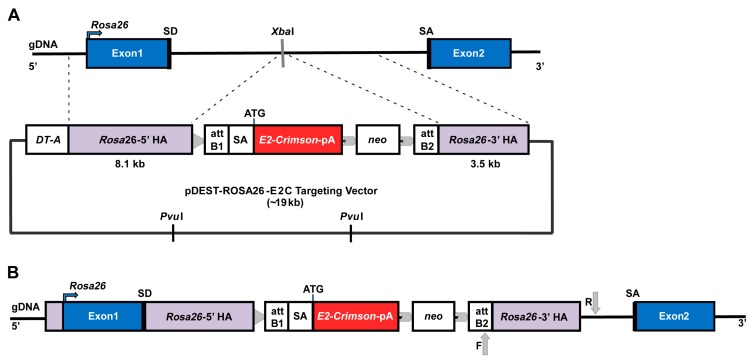
Figure 1.
Schematic illustration of the Rosa26 knock-in strategy. (A) E14-Bra-GFP mESC Rosa26 chromosome structure and pDest-ROSA26-E2C targeting vector. The vector contains two homology arms (HAs) that flank the E2-Crimson cassette, which has a polyadenylation (pA) signal, and a downstream positive drug selection marker neo (neomycin resistance). The insertion site in between the HAs is identified by the XbaI restriction enzyme site in the Rosa26 locus. A lethal negative selection marker DT-A is placed in the 5′ upstream region adjacent to the targeting arm. The PvuI restriction enzyme sites are located in the vector backbone. When linearised for gene targeting, most of the vector backbone is removed following PvuI restriction digestion; (B) E14-Bra-GFP mESC Rosa26 locus with correctly targeted insertion of linearised pDest-ROSA26-E2C targeting vector. In the correct recombinants, E2C cDNA is introduced into the Rosa26 locus and is expressed under the control of the Rosa26 promoter. A loxP site (indicated with a grey triangle) is located next to the 5′ HA. Flippase recombinase target (FRT) sites (indicated with grey arrow heads) flanking the neo cassette are designed for optional removal of the neo cassette. Grey arrows show the forward and reverse primer-binding sites for the PCR screen of correct homologous 3′-HA recombination.

