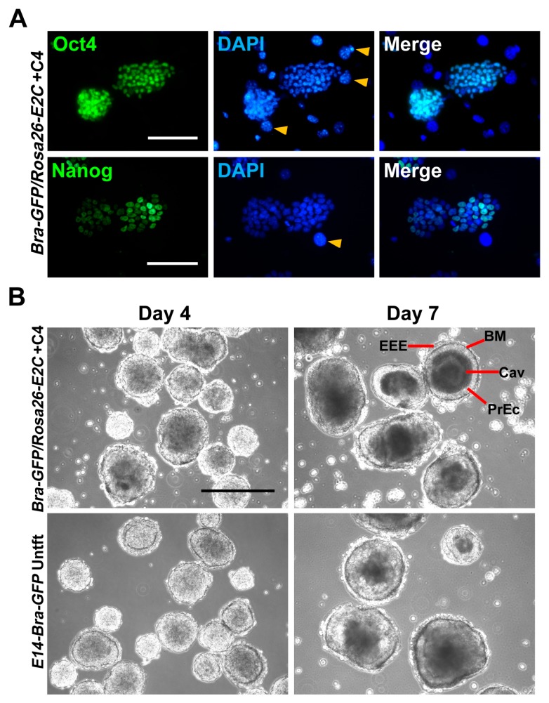
Figure 4.
(A) Expression of stemness markers Oct4 and Nanog was confirmed by immunofluorescent staining of Bra-GFP/Rosa26-E2C mESC clone +C4. All samples were counter-stained with DAPI. Yellow arrow heads show STO feeder cell nuclei; (B) Differentiation potential of clone +C4 was confirmed by typical EB formation. Cells were seeded in suspension culture dishes up to day 7. PrEc, primitive ectoderm; EEE, extraembryonic endoderm; BM, basement membrane; Cav, cavity. Scale bars, 100 µm (A); 400 µm (B).

