Figure 6.
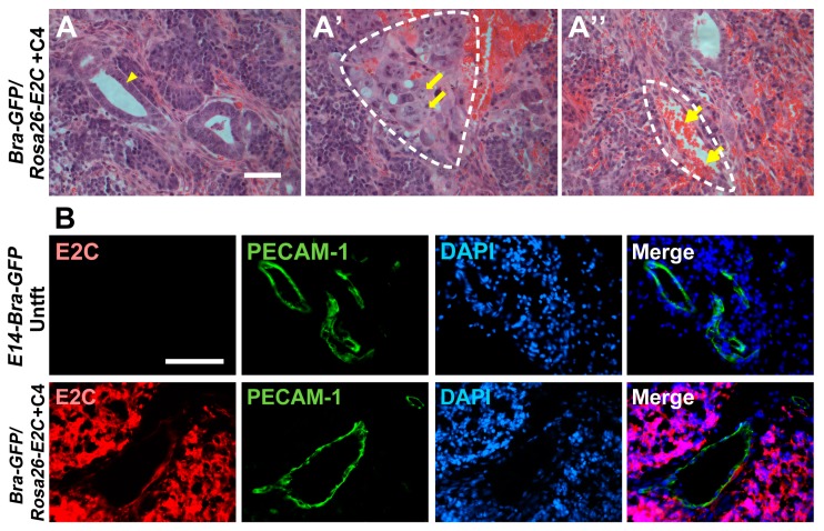
Histopathology and immunofluorescence analyses of tumours derived from the mESCs. (A–A′′) Haematoxylin and eosin (H&E) staining was performed on paraffin sections prepared from the day-9 tumours. Arrow heads show epithelial structure (A), thick arrows show chondrocyte-like cells (A′) and thin arrows show red blood cells (A′′). Dashed lines show the structures of chondrogenic-like (mesoderm-like) differentiation (A′) and blood vessels (A′′); (B) Immunostaining for E2C (red) and PECAM-1 (green) in tumours derived from Bra-GFP/Rosa26-E2C mESCs or untransfected controls (Untft). Dual immunostaining of frozen sections prepared from tumours harvested at day 9 showed that all cells, except the vasculature and blood cells, within the tumours were derived from the E2C+ mESC, which stained positively for E2C. On the other hand, the endothelial cells within the tumours did not stain positively for E2C, indicating they were derived from the host animals. Tumours developed from the untransfected E14-Bra-GFP mESCs were used as controls. Scale bars, 50 µm (A–A′′); 100 µm (B).

